MRI Use: Conditional to 7T
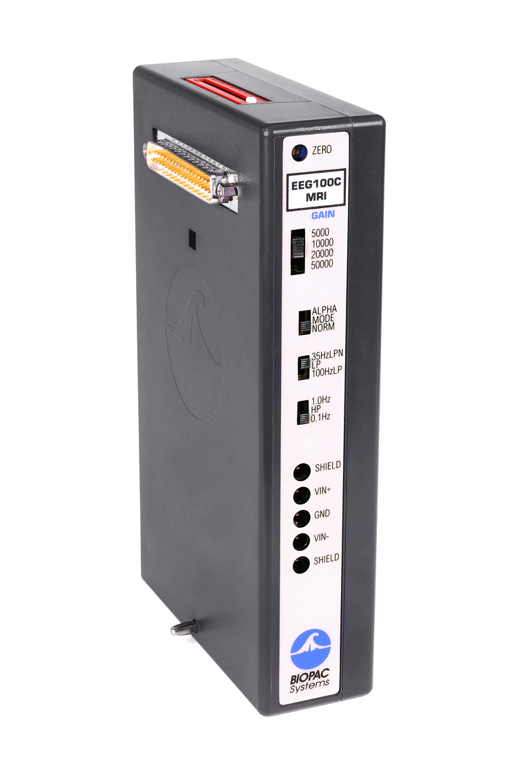
Condition: EEG100C-MRI Amplifier stays in the Control Room and is used with the MECMRI-BIOP filtered cable set and recommended MR leads/electrodes/transducers; tested to 7T.
IMPORTANT! See Safety Guidelines for recording biopotential measurements in the MRI environment.
Features:
- Less sensitivity to electrode and transducer lead placement
- Improved gain selectability
- No missing spectra in physiological signal frequency band
- Minimizes computer-based real-time or post-processing signal processing
- Cleaner data available as real-time analog output
The MRI Smart Amplifiers incorporate advanced signal processing circuitry which removes spurious MRI artifact from the source physiological data. Signal processors are able to distinguish between physiological signal and MRI artifact as manifested by gradient switching during MRI sequences, such as Shim or EPI.
Because MRI-related transient artifact is removed at the source, the MRI version amplifier can be sampled at the same rate as during normal (non-MRI) physiological recording. In every aspect, data recording is easier and the final results are cleaner when using the MRI version amplifiers to record physiological data in the fMRI or MRI.
Product Family
Product Options
Platform Options
MODULAR CONSTRUCTION
Amplifiers snap together for easy system configuration and re-configuration.
Intuitive, Elegant AcqKnowledge Software
Powerful automated analysis. Instantly & easily view, measure, analyze, transform, and report data.
Powerful MP160 Data Acquisition and Analysis System
Flexible, proven modular data acquisition and analysis system for life science research.



Stay Connected