Magnetic Resonance Imaging with BIOPAC Equipment
Record Physiological Signals During MRI
Options for human and small animal MRI life science research
Advanced line of MR Safe and MR Conditional solutions

High-Quality Data for Human & Animal Protocols
BIOPAC® offers a series of amplifiers, electrodes, electrode leads, transducers, and stimulus options for safe data acquisition of physiological signals in the MRI environment. Specialized cable systems provide isolated and RF filtered interfacing between the subject/chamber panel and the control room.
Try MRI tools in the free AcqKnowledge Demo
Record physiological signals such as: Electrocardiogram (ECG), Electromyogram (EMG), Electroculogram (EOG), Electrogastrogram (EGG), Temperature, Respiration, Electrodermal Activity (EDA, EDR, SCL, SCR or GSR), Pulse, Hand Grip Strength (Dynamometry), CO2 and O2 Gas Analysis, Pulse Oximeter (SpO2), Finger Twitch, and a variety of pressure based signals. Microvascular blood perfusion can be measured with the Laser Doppler Flow System. Electrical stimulation is also available for constant current and constant voltage electrical stimulation. Physiological signals in the MRI can be used for psychophysiological studies, neuromarketing, subject monitoring, and more.
See More...Hardware Packages | Magnetic Resonance Imaging with BIOPAC Equipment
Hardware Bundles are complete solutions for the specified application. Choose your preferred platform and bundle, then click "Request Pricing" to request an estimate, add/remove items, or complete purchase. If you have questions about specific items, click through to the product web page for details and specifications, or contact your Local Sales contact.
MRI
-
MP160 System + Setup for Facial EMG in MRI
MRI | Magnetic Resonance Imaging with BIOPAC Equipment
MP160 with AcqKnowledge plus EMG MRI amp with EL510 disposable electrode setup and impedance checker; includes 100 pre-wired 3-lead Ag-AgCl radio-translucent electrodes.
-
Dual-channel Gating System for MRI (ECG & Respiration)
MRI | Magnetic Resonance Imaging with BIOPAC Equipment
Gate System includes an MP160 data acquisition & analysis system with AcqKnowledge software and provides the cardiac trigger via an MRI Smart Electrocardiogram Amplifier (ECG100C-MRI), dual-channel cardiac respiratory gating system (DTU300 for human or large animal), MR Safe respiration transducer (TSD110-MRI), and cables, electrodes, and leads. Bundle also includes Impedance Checker and ELPAD.
-
MP160 System + ECG, EDA & Respiration for MRI
MRI | Magnetic Resonance Imaging with BIOPAC Equipment
MP160 with AcqKnowledge plus ECG, EDA & Respiration setups for MRI
MP160 System + Setup for Facial EMG in MRI
- 1 x MP160 Data Acquisition Systems
- 1 x EMG Electromyogram Amplifier for MRI
- 1 x MRI Filtered Cable Sets
- 5 x Disposable RT Electrodes - 3/set (pk of 20 sets)
- 1 x EEG/ECG/EMG/EOG Prep Gel 114 g
- 1 x Electrode Impedance Checker
Dual-channel Gating System for MRI (ECG & Respiration)
- 1 x Cardio Respiratory Gating System - ECG
- 1 x Electrode Impedance Checker
- 1 x Abrasive Pads 10/pk
MP160 System + ECG, EDA & Respiration for MRI
- 1 x MP160 Data Acquisition Systems
- 1 x MRI Filtered Cable Sets
- 1 x MRI Filtered Cable Sets
- 4 x Leads - RT for MRI
- 1 x Electrode Gels
- 1 x ECG Electrocardiogram Amplifier for MRI
- 1 x Disp. Radiotranslucent Electrode, 100/pk
- 1 x Abrasive Pads 10/pk
- 1 x Electrode Impedance Checker
- 1 x General Purpose Transducer Amplifier
- 1 x Respiration Transducer for MRI
- 1 x EDA Electrodermal Activity Amplifier for MRI
- 1 x Disp. RT Dry Electrode for EDA, 100/pk
- 1 x Electrode Gel, Isotonic, 114 g
Animal
-
Dual-channel Gating System for MRI (ECG & Respiration)
Animal | Magnetic Resonance Imaging with BIOPAC Equipment
Gate System includes an MP160 data acquisition & analysis system with AcqKnowledge software and provides the cardiac trigger via an MRI Smart Electrocardiogram Amplifier (ECG100C-MRI), dual-channel cardiac respiratory gating system (DTU300 for human or large animal), MR Safe respiration transducer (TSD110-MRI), and cables, electrodes, and leads. Bundle also includes Impedance Checker and ELPAD.
Dual-channel Gating System for MRI (ECG & Respiration)
- 1 x Cardio Respiratory Gating System - ECG
- 1 x Electrode Impedance Checker
- 1 x Abrasive Pads 10/pk
Details
MRI Signal Conditioning—Automated Routines

Run the Magnetic Resonance Imaging routines for automated signal conditioning to focus on the signal of interest by removing MR gradient and other interference. Use individual filter options from the Transform and Analysis menus to further refine data cleaning parameters.
AcqKnowledge Filters for Additional Signal Clean-up
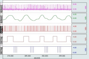
When subjects are monitored in the fMRI, imaging sequence types impact the physiological signal recording.
- In the case of Blood Oxygen Level Dependent (BOLD) imaging, T1 is unaffected by blood oxygen levels but T2 is greatly affected by blood oxygen levels. Impact varies based on the type of Blood Oxygen Level Dependent Effect (BOLD) Imaging, including
- Echo Planar Imaging (EPI): EPI is a magnetic resonance imaging (MRI) method valuable to neuroscience. EPI is employed for nearly all functional MRI (fMRI) and diffusion imaging of neural fiber connections in the brain.
- Multiband (MB) Excitation: MB excitation, during EPI, allows for the simultaneous acquisition of multiple slices during imaging. MB factor 6, means that six slices are acquired in the same time that one slice would be acquired during regular EPI. This technique is helpful for fMRI studies to improve the statistical definition of neural fiber connections and for diffusion-based imaging neural fiber tractography, to visualize structural connections in the brain.
- Axial EPI MB scans generate significant harmonics in the recorded physiological data at the point of scanning commencement; Coronal EPI MB scans generate significantly less harmonic energy.
- Multiplexed-EPI (M-EPI) Sequencing: M-EPI sequencing combines two forms of image acquisition multiplexing: time-based multiplexing (T) and space-based multiplexing (S) with Multiband RF pulses to acquire (T x S) images in an EPI sequence instead of just one image. M-EPI provides a substantial increase in time resolution and higher statistical power for fMRI, resulting in improved image resolution.
In these cases, the resulting scanning artifact can’t be removed by the MRI Smart Amplifiers because it falls close enough to the range and type of the physiological signal of interest. However, the data can be cleaned in AcqKnowledge by employing a comb-band-stop (CBS) filter to remove the offending harmonics, in post-processing or real time. The data will be well-scrubbed of artifact but some non-harmonic-related artifact presence will remain; this artifact is likely related to non-homogeneous qualities associated with the subject’s body and switching magnetic fields. Request Application Note 283 to learn more about fMRI Imaging Sequencing Effects upon Physiological Measurement.
QUALITY IMAGES WITH CARDIORESPIRATORY GATING SYSTEMS
Complete triggering and gating solutions for small animal and human subjects
MRI Smart Amplifiers
Advanced signal processing circuitry removes spurious MRI artifact from the source physiological data
Full line of specialized equipment
Transducers, cables/filters, amplifiers, electrodes & other accessories
Features & Benefits
Record and Monitor subjects in the scanner
- Human and Animal setups for fMRI
- MRI Smart Hardware for cleaner data
- Trigger Systems
- Gating Systems
- Remote Monitor (licensed feature)
- Plus more—Review the MRI Catalog
Automated Analysis Routines with AcqKnowledge
- MRI Signal Conditioning
Advanced Features Spotlight
Use the DTU100 Digital Trigger Unit to trigger an MRI System with the occurrence of the R-wave present in ECG, respiratory data or blood pressure for More...

Remote Monitor - IP-Based Network Display
Simplified view of subject data on another computer or mobile device. Remote Monitoring provides a simple browser interface, from which acquisitions can be started, stopped More...
The BIOPAC MP160 System supports small animal MRI monitoring system for ECG, Heart Rate, EMG, blood pressure, respiration, temperature, pulse oximetry, CO2 and O2 gas More...
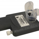
The TSD117-MRI is used outside the bore in the MRI Chamber Room and AFT7-L tubing is connected to reach the subject. Use coupler AFT11D to add More...

Record ECG, heart rate, and R-R interval from a subject inside a functional MRI with a BIOPAC MP150 data acquisition system. The AcqKnowledge software (included More...
Videos
Dual-Channel MRI Gating
MRI - Cleaning Data - EMG example
BIOPAC NIBP-MRI: How to Apply Sensor to Brachial Artery
Slew Rate Limiter in AcqKnowledge
Support
Application Notes
- 170 - Laser Doppler Flow Module - LDF100C
- 195 - MP System Data Sampling Reference
- 223 - Physiological Measurement in Magnetic Resonance Imaging Systems
- 121 - Waveform Data Reduction
- 142 - Rate Detector Algorithm in AcqKnowledge
- 148 - Automated ECG Analysis
- 182 - Analysis of Blood Flow Data
- 204 - AcqKnowledge Peak Detector Operation
- 211 - EEG Analysis With AcqKnowledge
- 118 - EMG Signal Analysis
- 165 - Integrated EMG
- 230 - Connections for Physiological Signals in an MRI
- 235 - Zygomaticus Measures with Pressure Pad vs. EMG in MRI or fMRI
- 001 - AcqKnowledge Release and OS Compatibility
- 241 - Recording EMG data in an fMRI
- 242 - Recording ECG Data in an fMRI
- 243 - Gated Analysis for Data Recorded in an MRI - EMG example
- 245 - DTU200 Dual Channel Gating Setup and Operating Instructions
- 258 - BIOPAC MRI Smart Amplifier Performance
- 259 - ECG Averaging in the MRI using the ECG100C-MRI amplifier
- 263 - Strategies to Record Biopotential or Transducer Data in MRI or fMRI
- 265 - MRI Artifact Removal from ECG Waveforms
- 279 - ECG and EDA Recording in 7-Tesla fMRI
- 281 - ECG Timing Delay Associated with Amplifier Filter Selections
- 282 - Subject Electrical Stimulation in fMRI and MRI
- 283 - fMRI Imaging Sequencing Effects upon Physiological Measurement
Knowledge Base
- * CLEANING GUIDELINES *
- Amplifier conversion - 2 mm to touchproof
- Audio recording synchronization
- Biopotential amplifier signal validation
- CAP100C electrode connections
- Common mistakes/general troubleshooting
- Copy and paste append markers along with data
- DA100C amplifier signal validation
- Duplicating waveforms
- ECG artifact in EMG Signal
- ECG R-wave detector
- EDA / SCR offset calibration for MRI
- Electrode Properties - gel and adhesive
- Exporting Data to SPSS
- External Devices and Channel Contention
- MRI Data Cleaning | PhysIO Toolbox to Remove Image Artifact
- MRI, Radiotranslucent, and Radio-opaque Compatibility
- Optimal ground placement
- Physiological measurements - life science signals using BIOPAC
- Text files - import and export
Stay Connected