Amplifier Modules
MRI Smart Amplifiers
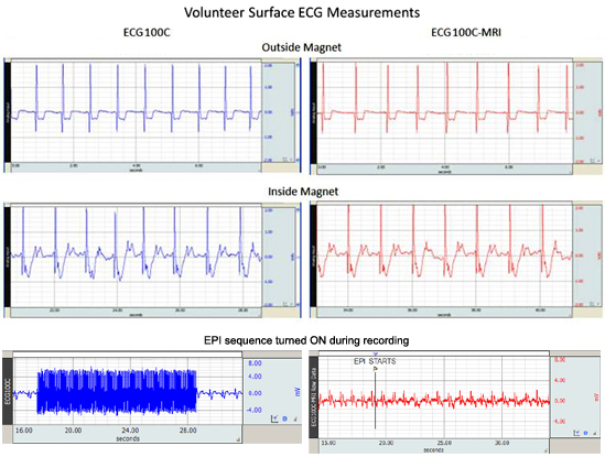
BIOPAC MRI Smart amplifiers incorporate advanced signal processing circuitry which removes spurious MRI artifact from the source physiological data.Signal processors are able to distinguish between physiological signal and MRI artifact as manifested by gradient switching during MRI sequences, such as Shim or EPI. BIOPAC has special expertise in research conducted in the MRI and provides a number of solutions for human and small animal MRI applications.
In every aspect, data recording is easier and the final results are cleaner when using the BIOPAC MRI version amplifiers to record physiological data in the fMRI or MRI.
Features
- Less sensitivity to electrode and transducer lead placement
- Improved gain selectability
- No missing spectra in physiological signal frequency band
- MRI-related transient artifact removed at the source
- Improved signal reduces the need for computer-based real-time or post-processing signal processing
- Cleaner data available as real-time analog output
1. Place electrodes on the subject according to these guidelines:
B) Attach the EL508 or EL509 electrodes as close to each other as possible, on the subject’s skin, for the measurement.
C) Place electrodes in as straight of a line as possible which is perpendicular to the magnet’s axis.
D) Place electrodes between 3-5 cm apart, if possible; the larger the area between the electrodes, the stronger the MRI gradient artifact.
2. Connect the electrode lead set to the electrodes according to these guidelines:
A ) Make sure that the electrode leads do not loop in a “circle”, “S” or “U” shape. Also, do not twist or braid the electrode leads. Looped, braided or twisted leads pick up RF energy, resulting in current induction and increased localized heating.
B ) Run the leads out of the chamber bore in the simplest (straightest) manner possible.
C) Do not allow the electrode leads to touch the subject’s bare skin. Electrode leads may heat up in the MRI.
Use a thermal insulator (such as a blanket or towel) between the electrode lead and the subject’s skin.
It’s also possible to use thermally-insulating foam jacket, similar to those used for insulating copper tubing, for placing the electrode leads to keep them away from the subject’s skin.
Stay Connected