BIOPAC® Systems, Inc. Logo

ECG Electrocardiogram Amplifier for MRI

Selectable signal conditioning—filter or transform data as it is being collected

Cascade up to 16 amplifer modules per MP Research System for multi-parameter or multi-subject studies

Request More Info
ECG Electrocardiogram MRI-Amplifier
Part #: ECG100C-MRI

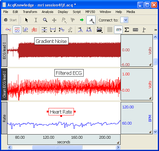
The ECG100C-MRI Electrocardiogram Amplifier records electrical activity generated by the heart and will reliably record ECG from humans, animals and isolated organ preparations. The amplifier output can be switched between normal ECG output and R-wave detection. The R-wave mode outputs a smoothed pulse with the occurrence of each R-wave. The exact timing of the R-wave is detected even under conditions of extreme signal artifact. The amplifier also includes a user-switchable baseline stabilizer.

Use the AcqKnowledge software to provide a complete Lead II ECG analysis. The software automatically scores the data and extracts the measurements of interest on a cycle-by-cycle basis. The results are automatically exported to Excel or pasted in the Journal file. AcqKnowledge also includes a fully automated HRV analysis feature. The HRV analysis provides values for VLF, LF, HF, VHF, sympathetic, and vagal, as well as the sympathetic / vagal balance.

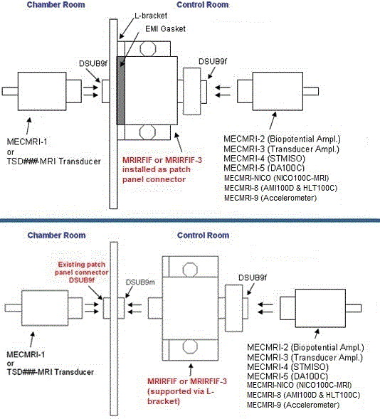
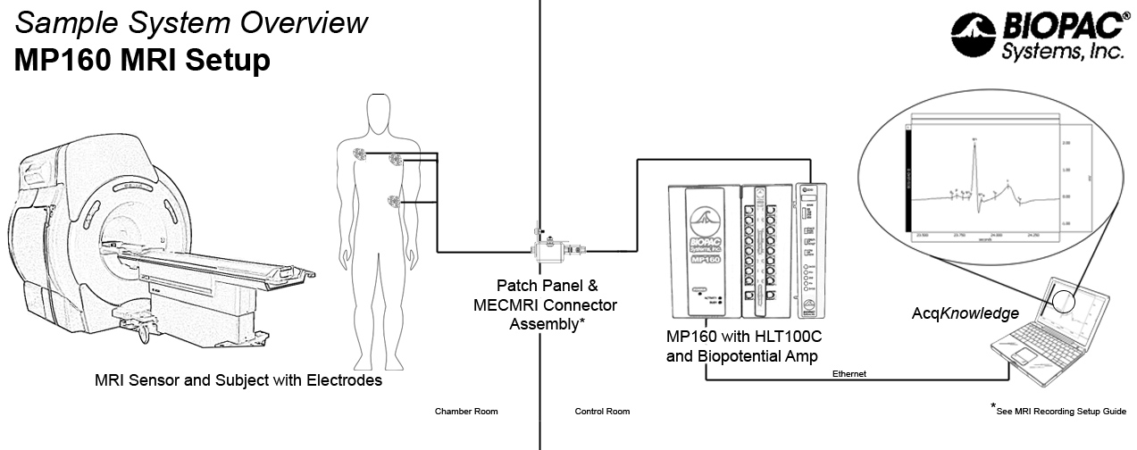
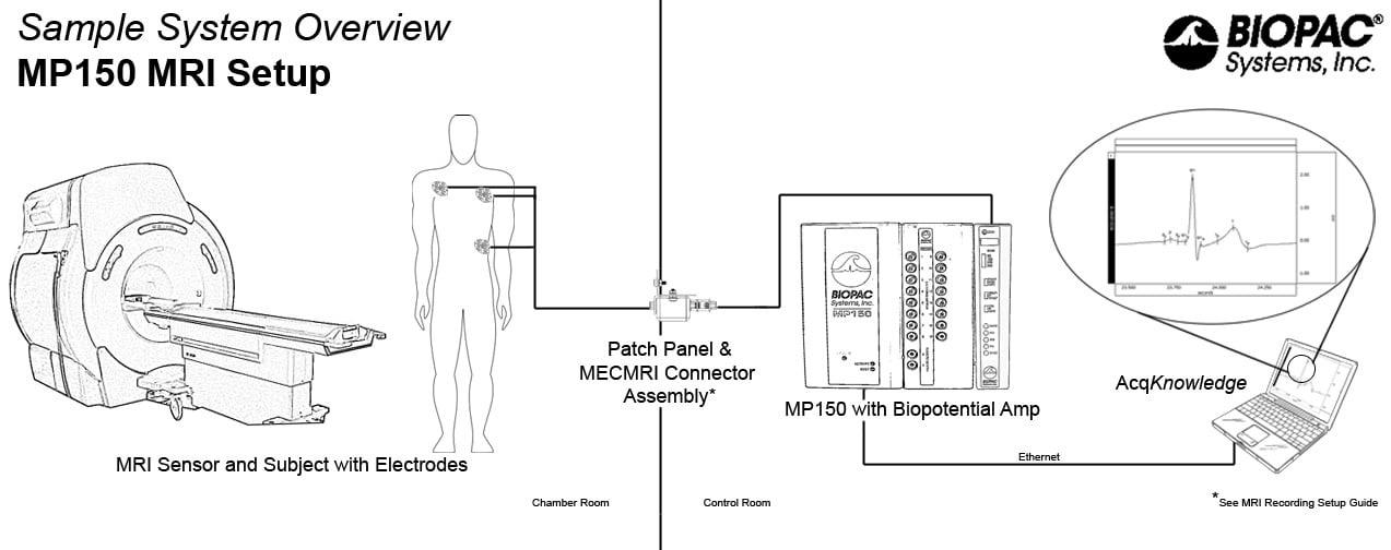
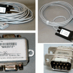
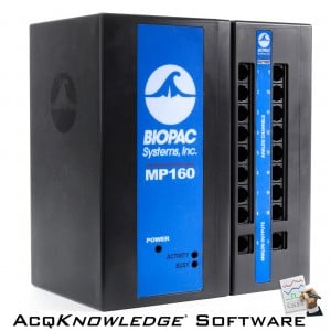
The ECG100C-MRI amplifier should be used with the MECMRI-BIOP MRI cable/filter set. The ECG-MRI is part of a complete research system, interfacing with the MP160/MP150 data acquisition and analysis platform and AcqKnowledge software, allowing advanced analysis for multiple applications and supporting acquisition of a broad range of signals and measurements.

See More...

MODULAR CONSTRUCTION

Amplifiers snap together for easy system configuration and re-configuration.

Intuitive, Elegant AcqKnowledge Software

Powerful automated analysis. Instantly & easily view, measure, analyze, transform, and report data.

Powerful MP160 Data Acquisition and Analysis System

Flexible, proven modular data acquisition and analysis system for life science research.

BIOPAC data acquisition
View Spec PDF

Are You Looking for These?

Part #: ECG100C-MRI
Categories: Amplifiers - Research, MRI Magnetic Resonance Imaging - Research
Subcategories: Biopotential Amplifiers - Research, Amplifier Modules - Research

Alternatives
For the same Platform

Equivalents
With a different Platform

Details

Compatibility


MRI Use: Conditional to 7T

Condition: ECG100C-MRI Amplifier stays in the Control Room and is used with the MECMRI-BIOP filtered cable set and recommended MR leads/electrodes/transducers; tested to 7T.

Usage Notes for MRI Smart Amplifiers

IMPORTANT! See Safety Guidelines for recording biopotential measurements in the MRI environment.

IMPORTANT! BIOPAC recommends using the shortest possible electrode leads for recording in the fMRI or MRI.

12-lead ECG Options

  • For full, simultaneous, 12-lead ECG recording, use a total of eight ECG100C-MRI amplifiers and a WT100C Wilson terminal (virtual reference). Use two ECG100C-MRI to record Leads I and II, from which the software will calculate Lead III, aVR, aVL and aVF, and use six ECG100C-MRI to simultaneously generate the six precordial chest leads (V1-V6).

MRI/CT Scan Trigger Option

  • To trigger an MRI System or CT Scanner with the occurrence of the R-wave present in animal (high frequency) ECG or respiratory data for gating purposes, use the DTU100 Digital Trigger Unit or the DTU200 Gating System.

MRI Smart Amplifier Features

In every aspect, data recording is easier and final results are cleaner when MRI Smart Amplifiers are used to record physiological data in the fMRI or MRI.

  1. Less sensitivity to electrode and transducer lead placement
  2. Improved gain selectability
  3. No missing spectra in physiological signal frequency band
  4. Minimizes computer-based real-time or post-processing signal processing
  5. Cleaner data available as real-time analog output

MRI Smart Amplifiers incorporate advanced signal processing circuitry which removes spurious MRI artifact from the source physiological data. Signal processors are able to distinguish between physiological signal and MRI artifact as manifested by gradient switching during MRI sequences, such as Shim or EPI. Because MRI-related transient artifact is removed at the source, the MRI version amplifier can be sampled at the same rate as during normal (non-MRI) physiological recording.

Recommended Reading

Thoralf Niendorf, Lukas Winter and Tobias Frauenrath (2012). Electrocardiogram in an MRI Environment: Clinical Needs, Practical Considerations, Safety Implications, Technical Solutions and Future Directions, Advances in Electrocardiograms – Methods and Analysis, PhD. Richard Millis (Ed.), ISBN: 978-953-307-923-3, InTech, DOI: 10.5772/24340.

Thakor NV, Webster JG, Tompkins WJ: Estimation of QRS Complex Power Spectra for Design of a QRS FilterIEEE Transactions on Biomedical Engineering 1984, 31(11):702-706. Abstract.

  • This is an algorithmic method to extract the R-Wave timing from a possibly corrupted ECG, or one where the R-wave is suppressed. BIOPAC R-wave detectors in the ECG100C and ECG100C-MRI both employ this strategy for detecting R-waves, as an option.

Featured Citation: ECG and ICG during BOLD fMRI

A study performed BOLD fMRI while simultaneously recording the Electrocardiogram (ECG) and Impedance Cardiogram (ICG) with BIOPAC MRI Smart Amplifiers and accessories. The resolution of the ECG and ICG waveforms was sufficient for measuring stroke volume, cardiac output, pre-ejection period, and left ventricular ejection time during imaging. Results indicate that ICG can be recorded during EPI and, with specific signal processing, can be interpreted similarly as to when recorded outside the MRI. Read more about the technique and results at Psychophysiology:

Cieslak, M., Ryan, W. S., Macy, A., Kelsey, R. M., Cornick, J. E., Verket, M., Blascovich, J. and Grafton, S. (2014), Simultaneous acquisition of functional magnetic resonance images and impedance cardiography. Psychophysiology. doi: 10.1111/psyp.12385

Videos

BIOPAC Research Solutions | ECG

Automated ECG Analysis- Lead II

HRV Analysis: Fully-automated Routine in AcqKnowledge

Support

Application Notes

Applications

Downloads/Resources

Knowledge Base

Recommended Items

Spotlight On
Cardiac Output amplifier

NICO Smart Amplifier

The NICO Smart Amplifier is specifically designed to measure thoracic impedance for performing noninvasive cardiac output measurements. The amplifier terminates in two connectors for impedance (Z) and its derivative (dZ), and has a 3 m cable that connects directly to an AMI100D Amplifier Input Module. Use with EL526 strip electrodes and CBL246 adapter or with EL500 […]

View All
Latest News

New Citations | BIOPAC in ANS Research

The autonomic nervous system (ANS) is responsible for regulating the functions of all the body’s organs. Take a look at this month’s citations to see how to apply BIOPAC’s tools and learn techniques for ANS research… Comparable responses to a wide range of olfactory stimulation in women and men. Scientific Reports, 13(1), 9059. Lillqvist, M., Claeson, […]

Electrocardiography Guide Available

BIOPAC’s comprehensive Introductory ECG Guide addresses fundamental to advanced concerns to optimize electrocardiography data recording and analysis. Topics include: ECG Complex; Electrical and Mechanical Sequence of a Heartbeat; Systole and Diastole; Configurations for Lead I, Lead II, Lead III, 6-lead ECG, 12-lead ECG, precordial leads; Ventricular Late Potentials (VLPs); ECG Measurement Tools; Automated Analysis Routines for extracting, […]

Read All
Request a Demonstration