

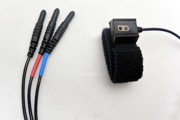
The transducer optics are designed to sense diffuse surfaces, including the skin surfaces of finger or toe. The transducer is sensitive to Blood Volume Pulse (BVP) via photo-plethysmographic methods.
The Diode and Phototransistor are mounted side by side on parallel axis in a black polyurethane housing. The Phototransistor is encased in a dark epoxy package which filters out visible ambient light. The transducer has a shielded 3-meter cable.
The ergonomic housing design improves contact with the subject and helps reduce motion artifact.
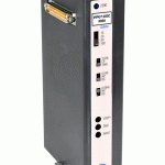
The TSD200-MRI only operates with the PPG100C-MRI amplifier.
Connections
- For MRI applications: Use the MECMRI-TRANS Cable/Filter to connect the TSD200-MRI to the PPG100C-MRI. See BIOPAC Application Notes regarding the proper installation of MECMRI cables for recording in an MRI environment.
- For non-MRI applications: Connect the TSD200-MRI directly to the PPG100C-MRI.
MRI Use: Conditional to 7T
Condition: Conductive parts of transducer are electrically and thermally isolated from subject. (See Specifications for components.)
Spotlight Application
Autonomic arousals contribute to brain fluid pulsations during sleep
Dante Picchioni, Pinar S. Özbay, Hendrik Mandelkow, Jacco A. de Zwart, Yicun Wang, Peter van Gelderen, Jeff H. Duyn,
NeuroImage, Volume 249, 2022, 118888, ISSN 1053-8119, https://doi.org/10.1016/j.neuroimage.2022.118888.
(https://www.sciencedirect.com/science/article/pii/S1053811922000180)
“Concurrently acquired peripheral physiological measures included chest belt (to monitor respiratory chest excursion) and finger skin photoplethysmography (PPG) (to monitor cardiac rate and peripheral vascular volume). These were acquired using a BIOPAC acquisition system with TSD200-MRI and TSD221-MRI transducers, and an MP150 digitizer sampling at 1 kHz (BIOPAC, Goleta, CA, USA). Data collection for both EEG and peripheral physiology were synchronized with the fMRI through a trigger signal provided by the MRI scanner.”
Stay Connected