BIOPAC® Systems, Inc. Logo

Respiration Transducer for MRI

Transducer for MRI Applications

Record responses in the MRI

Request More Info
Respiration Transducer for MRI
Part #: TSD221-MRI

This MR Safe fully-pneumatic respiration transducer measures subject respiration (thoracic or abdominal) in the MRI. The extremely unobtrusive design presents minimal resistance to movement and can measure arbitrarily slow to very fast respiration patterns with no loss in signal amplitude, while maintaining excellent linearity and minimal hysteresis.

The TSD221-MRI includes the respiration sensor in a mesh strap with self-adhering band (70 cm), a pressure transducer (±2.5 cm H2O TSD160A), and cascadable tubing for up to 14 m (AFT30-XL 10 m + AFT30-L 4 m).

Connects to the DA100C and an MP160, MP150, or MP100 System—does not require cable set MECMRI-DA or MECMRI-TRANS.

MRI Use: MR Safe—TSD221-MRI has no ferrous metals or conductive parts (see Specifications for components).

See More...

Respiratory Effort Transducer - MRI Safe
View Spec PDF

Are You Looking for These?

Part #: TSD221-MRI
Categories: MRI Magnetic Resonance Imaging - Research
Subcategories: Transducers - Research

Equivalents
With a different Platform

Details

Compatibility


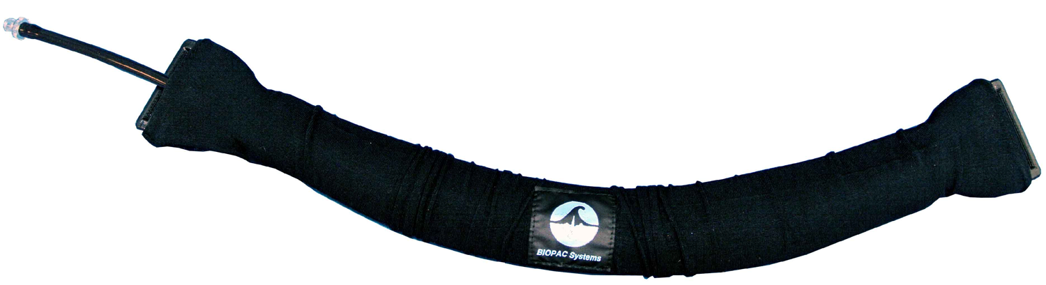
The TSD221-MRI is very easy to set up, in the MRI, due to its fully MR Safe design.

  • Place the respiration transducer around the body at the level of maximum respiratory expansion, generally about 5 cm below the armpits but location will vary from the erect to supine positions.
    • To obtain proper tension, stretch the belt around the body and have the subject exhale. At maximum expiration, adjust the nylon strap so there is slight tension to hold the strap around the chest.Correct tension adjustment of the respiration transducer is important. For best sensitivity, the transducer must be just slightly tight at the point of minimum circumference (maximum expiration).
  • The coupling pneumatic tube connects to the respiration sensor on the subject and should be directed through an available waveguide to terminate on the TSD160A pressure transducer attached to the DA100C in the Control Room. Measurement delay due to tubing length is 3 ms/meter.

Spotlight Application

Autonomic arousals contribute to brain fluid pulsations during sleep
Dante Picchioni, Pinar S. Özbay, Hendrik Mandelkow, Jacco A. de Zwart, Yicun Wang, Peter van Gelderen, Jeff H. Duyn,
NeuroImage, Volume 249, 2022, 118888, ISSN 1053-8119, https://doi.org/10.1016/j.neuroimage.2022.118888.
(https://www.sciencedirect.com/science/article/pii/S1053811922000180)

“Concurrently acquired peripheral physiological measures included chest belt (to monitor respiratory chest excursion) and finger skin photoplethysmography (PPG) (to monitor cardiac rate and peripheral vascular volume). These were acquired using a BIOPAC acquisition system with TSD200-MRI and TSD221-MRI transducers, and an MP150 digitizer sampling at 1 kHz (BIOPAC, Goleta, CA, USA). Data collection for both EEG and peripheral physiology were synchronized with the fMRI through a trigger signal provided by the MRI scanner.”


Support

Downloads/Resources

Knowledge Base

Recommended Items

Spotlight On
BSL Practical Lab Kit

Biopac Student Lab Human Physiology Systems

BSL System with MP36 and hardware for 11 guided lessons plus 14 dynamic lessons and student-designed Human Physiology experiments. Use the power of the BSL Human Physiology Teaching System to conduct the most widely studied responses in physiology teaching labs. Lessons target the circulatory system, respiratory system, muscular function, brain function, ANS, exercise physiology and […]

View All
Latest News

New Citations | BIOPAC in ANS Research

The autonomic nervous system (ANS) is responsible for regulating the functions of all the body’s organs. Take a look at this month’s citations to see how to apply BIOPAC’s tools and learn techniques for ANS research… Comparable responses to a wide range of olfactory stimulation in women and men. Scientific Reports, 13(1), 9059. Lillqvist, M., Claeson, […]

Electrocardiography Guide Available

BIOPAC’s comprehensive Introductory ECG Guide addresses fundamental to advanced concerns to optimize electrocardiography data recording and analysis. Topics include: ECG Complex; Electrical and Mechanical Sequence of a Heartbeat; Systole and Diastole; Configurations for Lead I, Lead II, Lead III, 6-lead ECG, 12-lead ECG, precordial leads; Ventricular Late Potentials (VLPs); ECG Measurement Tools; Automated Analysis Routines for extracting, […]

Read All
Request a Demonstration