

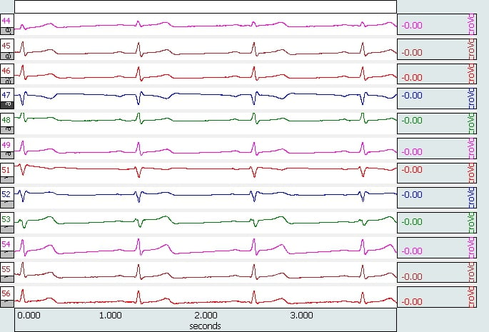
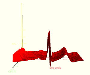
Use the AcqKnowledge software to provide a complete Lead II ECG analysis. The software automatically scores the data and extracts the measurements of interest on a cycle-by-cycle basis. The results are automatically exported to Excel or pasted in the Journal file. AcqKnowledge also includes a fully automated HRV analysis feature. The HRV analysis provides values for VLF, LF, HF, VHF, sympathetic, and vagal, as well as the sympathetic / vagal balance.
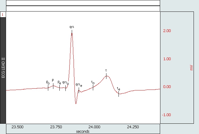
ECG Analysis Lead II
12-lead ECG Options
- For full, simultaneous, 12-lead ECG recording, use a total of eight ECG100C amplifiers and a WT100C Wilson terminal (virtual reference). Use two ECG100C to record Leads I and II, from which the software will calculate Lead III, aVR, aVL and aVF, and use six ECG100C to simultaneously generate the six precordial chest leads (V1-V6).
- To generate a 12-lead recording using seven simultaneous leads with a single precordial chest lead appended in data segments, use three ECG100C amplifiers and a TSD155C multi-lead ECG cable (which incorporates a built-in Wilson Terminal). Use two ECG100C to generate Leads I and II, from which the software will calculate Lead III, aVR, aVL and aVF, and use the other ECG100C with the TSD155C to record one (movable) precordial lead [V1, V2, V3, V4, V5 or V6] in appended data segments.
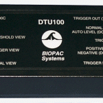
- To trigger an MRI System or CT Scanner with the occurrence of the R-wave present in animal (high frequency) ECG or respiratory data for gating purposes, use the DTU100 Digital Trigger Unit.
Recommended Reading
Thakor NV, Webster JG, Tompkins WJ: Estimation of QRS Complex Power Spectra for Design of a QRS Filter. IEEE Transactions on Biomedical Engineering 1984, 31(11):702-706. Abstract.
- This is an algorithmic method to extract the R-Wave timing from a possibly corrupted ECG, or one where the R-wave is suppressed. BIOPAC R-wave detectors in the ECG100C and ECG100C-MRI both employ this strategy for detecting R-waves, as an option.
Product Family
Product Type
Product Options
Platform Options
MODULAR CONSTRUCTION
Amplifiers snap together for easy system configuration and re-configuration.
Intuitive, Elegant AcqKnowledge Software
Powerful automated analysis. Instantly & easily view, measure, analyze, transform, and report data.
Powerful MP160 Data Acquisition and Analysis System
Flexible, proven modular data acquisition and analysis system for life science research.

Stay Connected